UChicago researchers identify neural circuit implicated in fear suppression

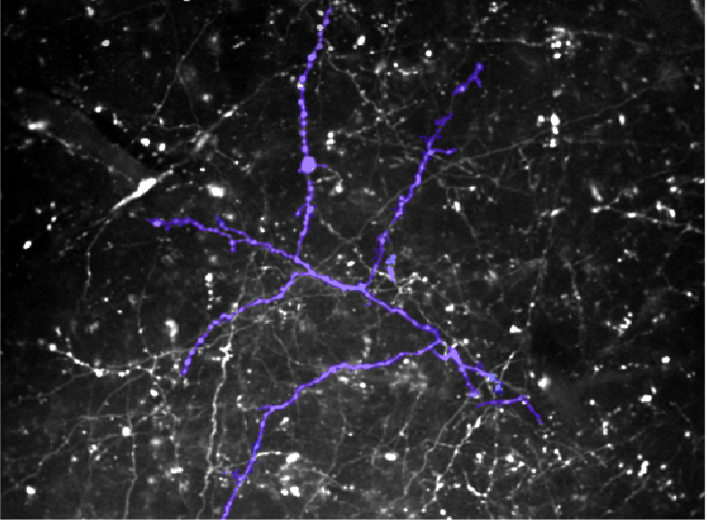
Single branching Nucleus Reuniens axon (highlighted in purple) in the CA1 region of the hippocampus. (Photo by Mark Sheffield)
Picture driving to work on a rainy day when suddenly you end up in a minor fender-bender. A quick repair and you’re back on the road, commuting daily as normal. One day, a light drizzle begins. You turn onto that familiar road and find yourself with sweaty palms and a racing heart. Ideally, this feeling fades after a few days of driving without incident. However, long-term, unwavering fear in response to a rainy drive would be detrimental to your mental health and lifestyle.
While contextual fear — fear tied to a specific situation or environment — is a crucial survival mechanism, suppressing fear is just as critical. Failure to do so underlies conditions such as generalized anxiety and post-traumatic stress disorder (PTSD). Regions of the brain responsible for encoding, retrieving, and suppressing fearful events are well studied, yet the specific circuits mediating this have remained elusive up until now. In a new study, University of Chicago neurobiology researchers have characterized the action of a fundamental pathway in contextual fear memory suppression.
“The brain is really good at making fearful memories,” said senior author Mark Sheffield, PhD, Assistant Professor of Neurobiology at UChicago. “When someone has an inability to suppress a contextual fear memory, they can't learn that their environment is safe. They're trapped— they are fearful when they shouldn't be. That's a big problem.”
Members of the Sheffield lab study mechanisms of memory using mouse models. In their new study, published October 24, 2023, in Nature Communications, they monitored live brain activity to demonstrate that a neural circuit bridging regions of the brain suppresses fear by disrupting the process by which fear memories are retrieved. This pathway also reduces inappropriate fear responses to non-dangerous situations and speeds up the rate of “extinction learning,” or the process by which fear fades away.
“I was not even aware that that circuit existed”, said Sheffield. “There was some literature showing that it’s anatomically there. People have imaged the brain and said, ‘Okay, there’s this connection there.’ But it hadn’t been studied.”
The neural path to fear suppression
While contextual fear memory is known to rely on the hippocampus, the role of other brain components in suppressing fear memory retrieval is more obscure. One candidate is the nucleus reuniens (NR), a subregion of the thalamus and a major communication hub with roles in emotional regulation and memory. The NR has input to the hippocampus via a subregion called CA1. The new study’s first author and Sheffield lab member Heather Ratigan, PhD, hypothesized that signals sent across this NR-CA1 connection suppress fear responses and encourage extinction learning.
This pathway suppresses the fear memory in real time, which allows the extinction learning to take place.
Mark Sheffield, PhD
To induce contextual fear memory, Ratigan and others designed a virtual reality (VR) program that was viewed by mice while they moved on a treadmill. Different VR “contexts” — simulated rooms with distinct combinations of lights, colors, and shapes — were paired with mild physical shocks to the tail. When re-exposed to these contexts in the absence of the shocks, the mice would freeze in place for short instances. The length and frequency of freezing instances provided researchers with an easily measurable readout of fear.
To draw connections of mouse behavior to activity in the brain, Ratigan inhibited some of the NR-CA1 projecting neurons during instances of fear. She found that mice began freezing longer and much more frequently, even when they weren’t viewing the shapes and colors in which they previously received the tail shocks. Using two-photon imaging, a technique used to visualize brain activity in real time, the researchers also observed that the activity of NR axons ramp up during context-dependent freezing episodes.
Together, Sheffield said, the results describe a role of NR-CA1 in fear extinction. “This pathway suppresses the fear memory in real time, which allows the extinction learning to take place. If you don't suppress the memory, you can't learn it's safe because you're just constantly retrieving the fearful part of the memory. You can't break out. That makes sense.”
Expanding the search
Looking ahead, Sheffield plans to turn his attention to the hippocampus by measuring the activity of the thousands of neurons receiving the NR-CA1 signal. Another idea is to enhance NR-CA1 activity instead of inhibiting it — an experiment with potential therapeutic significance for disorders such as PTSD that are characterized by persistent fear responses.
“Instead of shutting this circuit off, now we're going to boost it — we're going to make it hyperactive,” said Sheffield. “Can we shut down fear memories quicker, and cause that extinction learning to happen quicker? That has some real clinical relevance.”
Research reported in this press release was supported by The Whitehall Foundation, The Searle Scholars Program, The Sloan Foundation, The University of Chicago Institute for Neuroscience start-up funds, a New Innovator grant from the National Institutes of Health (1DP2NS111657-01) awarded to Mark Sheffield, and a T32 training grant (T32DA043469) from National Institute on Drug Abuse awarded to Seetha Krishnan.
This article was originally published on the BSD News website.